facs flow cytometry protocol
EdU 5-ethynyl-2-deoxyuridine is a nucleoside analog to thymidine and is incorporated into DNA. Add 01-10 μgml of the primary labeled antibody.
Overview Of Flow Cytometry Cell Signaling Technology
Collect cells by centrifugation and aspirate supernatant.

. Harvest wash the cells single cell suspension and adjust cell number to a concentration of 1-5106 cellsml in ice. Incubate on ice for 5 minutes. Wash the cells once with cold PBS at 300-400 x g and re-suspend.
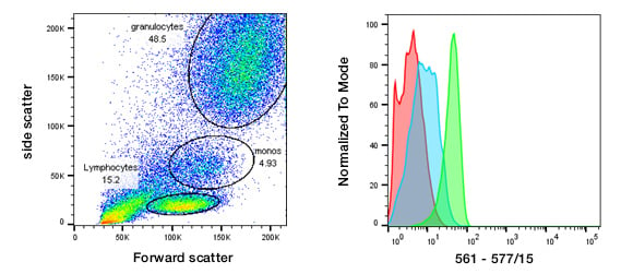
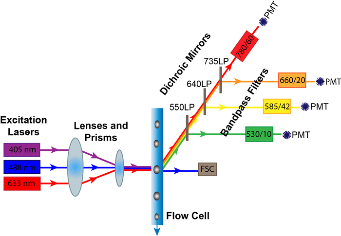
Perform red blood cell lysis per lab protocol either ACT ACK or LSM. Use this buffer also for all washes until directed to use Sorting Buffer Adjust. Flow cytometry FCM is a means of measuring certain physical and chemical characteristics of cells or particles as they pass in a.
Dilutions if necessary should be made in FACS buffer. The following flow cytometry. The Intacellular Flow Cytometry Staining Protocol describes the process for intracellular staining of various cell types in vivo-stimulated tissues in vitro-stimulated cultures and whole blood.
Flow Cytometry FCM and FACS protocols. Incubate for at least 30 min at room temperature or 4C in the dark. Stop cell lysis by adding 10ml Cell Staining Buffer to the tube.
Flow Cytometry is used for research applications such as immunophenotyping DNA studies cell cycle analysis and fluorescence-activated cell sorting FACS. Resuspend the cells to approximately 1-5 x 10 6 cellsml in ice cold PBS 10 FCS 1. Add the specific secondary antibodies at the proper dilution and incubate the cells at 4C on ice for 30 minutes in the dark.
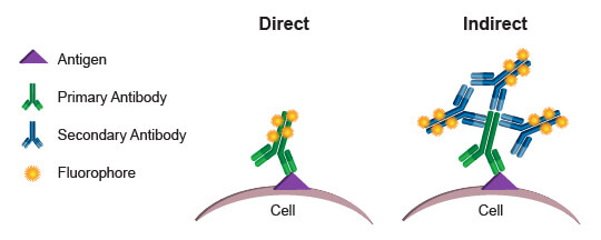
Anti-Neu5Gc Antibody Kit Protocol. By utilizing highly specific antibodies labeled with fluorescent conjugates FACS analysis allows us to simultaneously collect data on and sort a biological sample by a nearly limitless number. Cell Surface Flow Cytometry Staining Protocol.
Direct staining of cells applicable where the fluorophore. Flow cytometry FACS staining protocol Cell surface staining 1. It is always useful to check the viability of the cells which should be around 95 and not less than 90.
Repeat wash as in step 2. Precision Count Beads Protocol and Applications. Centrifuge for 5 minutes at 350xg and discard supernatant.
The flow cytometry protocols below provide detailed procedures for the treatment and staining of cells prior to using a flow cytometer. The Click-iT EdU Flow Cytometry Assay Kits are novel alternatives to the BrdU assay. Immunofluorescent Staining of Intracellular Cytokines for Flow Cytometric Analysis.
Flow cytometry and FACS fluorescence activated cell sorting are distinctly different procedures though FACS is a descendant procedure based upon flow cytometry. Please refer to the product webpage and product-specific protocol to determine whether it is compatible with live cell staining. Re-suspend in FACS staining buffer.
Get information on stimulation of cells appropriate cultures for generating human mouse and rat.
Controls For Flow Cytometry Bio Rad

Flow Cytometry Facs Protocols Sino Biological

Pdf How Is Flow Cytometry Protocol For Staining Intracellular Molecules Used Facs Analysis Academia Edu

Flow Cytometry Guide

Isolation Of Mouse Kidney Resident Cd8 T Cells For Flow Cytometry Analysis Protocol

Flow Cytometry Basics Facs Priciple How Does Flow Cytometry Work
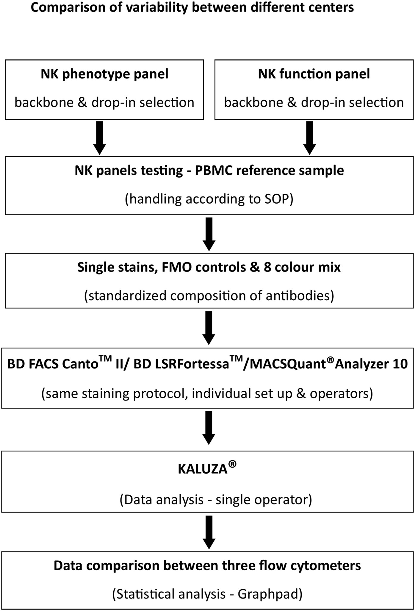
Standardized And Flexible Eight Colour Flow Cytometry Panels Harmonized Between Different Laboratories To Study Human Nk Cell Phenotype And Function Scientific Reports

Flow Cytometry Protocols For Surface And Intracellular Antigen Analyses Of Neural Cell Types Protocol

Workflow For Establishing The Fit For Purpose Of A Flow Cytometry Download Scientific Diagram
Development Application And Computational Analysis Of High Dimensional Fluorescent Antibody Panels For Single Cell Flow Cytometry Nature Protocols

Bacteria Counting And Enumeration Assays For Flow Cytometry Thermo Fisher Scientific Us
Stepwise Optimization Of The Procedure For Assessment Of Circulating Progenitor Cells In Patients With Myocardial Infarction Plos One

Flow Cytometry Detection Of Surface And Intracellular Antigens In Pancreas From A Single Mouse Embryo Star Protocols
Flow Cytometry Fc Protocol Epigentek
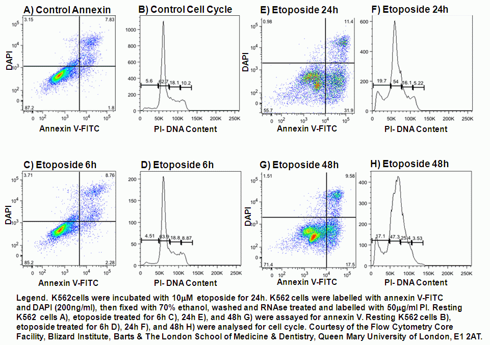
Annexin V And Apoptosis Flow Cytometry Core Facility

Immunophenotyping Hscs From Mouse Bone Marrow Protocol Deutschland

Sample Preparation For Flow Cytometry Best Practices
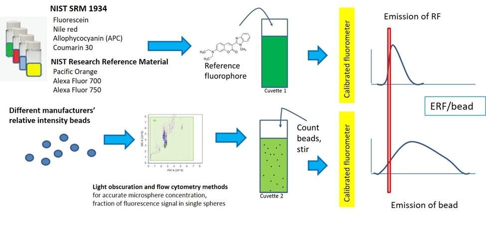
Quantitative Flow Cytometry Measurements Nist
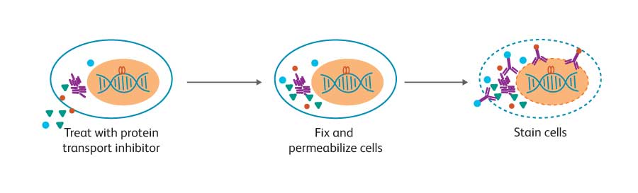
Intracellular Flow Cytometry Intracellular Staining